Meet Our Reporters
Imanis offers a wide-variety of reporters for noninvasive in vivo imaging. With optical, nuclear, and photoacoustic reporters, we’re sure you will find a reporter that suits your study needs.
Luciferase (bioluminescent imaging)
Near-infrared fluorescent protein (iRFP)
Tyrosinase (photoacoustic imaging)
Not sure which of our many reporters to choose? Check out our recommendations on choosing a reporter.
Nuclear Reporters
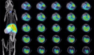
Nuclear reporters are proteins that trap various radiotracers in the cells in which they are expressed. Most nuclear reporters, including the sodium iodide symporter (NIS), human norepinephrine transporter (hNET), human somatostatin receptor 2 (hSSTR2), and human dopamine receptor D2 (hDRD2), are transporters or receptors that directly mediate uptake of specific radiotracers into the cells. These reporter proteins are endogenously expressed in various human tissues, where they have a diverse range of important functions. Another nuclear reporter, Herpes Simplex Virus Thymidine Kinase (HSVTK), is an enzyme that catalyzes the phosphorylation of intracellular radiolabeled pro-drugs so that they are no longer membrane permeable and are trapped in the cell. For imaging studies, target cells and viruses can be engineered to express high levels of any of the recombinant nuclear reporter proteins.
Because they concentrate radiotracers, nuclear reporters can be used to noninvasively track cells in vivo through nuclear imaging. Imaging with nuclear reporters offers several key advantages: it produces high-resolution 3D images that are fully quantitative, it is translational from small to large animals as well as humans (depending on the reporter), and it can be used for longitudinal imaging studies as species-specific versions exist for some of the reporters.
Check out our Discover NIS page for more detailed information about our favorite reporter.
How to use nuclear reporters for imaging
Reagents: Nuclear reporters trap a variety of different radiotracers. The following table indicates which radiotracers are compatible with each reporter.
|
Reporter |
SPECT radiotracers |
PET radiotracers |
|---|---|---|
|
NIS |
Iodine-123 Iodine-125 [99mTc]-pertechnetate |
Iodine-124 [18F]-TFB |
|
hNET |
[123I]-MIBG [125I]-MIBG |
[124I]-MIBG [11C]-ephedrine [11C]-mHED |
|
hSSTR2 |
[111In]-DTPA-octreotide [111In]-pentetreotide |
[68Ga]-DOTA-TATE [18F]-FP-Gluc-TOCA |
|
HSVTK |
[123I]-FIAU [125I]-FIAU |
[124I]-FIAU [11C]-FIAU [18F]-FIAU [18F]-FHBG |
|
hDRD2 |
[123I]-Ioflupane
|
[18F]-FESP [18F]-Fallypride |
Equipment: SPECT or PET machines can be used to detect radiotracer signal and biodistribution of nuclear reporter-expressing cells or viruses. Combining SPECT or PET with computed tomography (CT) or magnetic resonance (MR) imaging gives the most informative results, as images can be co-registered to give anatomical localization of the radiotracer signal.
Additional considerations: Most of the nuclear reporters are endogenously expressed in certain organs or tissues. When selecting a nuclear reporter, the imaging target site should be considered. Additionally, not all radiotracers may effectively reach certain target tissues/organs. See the chart under the “When to use nuclear reporters” section, below, for more information.
Workflow:

When to use nuclear reporters
Nuclear reporters are well suited for longitudinal, noninvasive imaging studies in regenerative medicine, gene therapy, oncology, and oncolytic virotherapy, where they can be used to track the biodistribution of cell/virus therapies or the destruction of tumors. Nuclear reporters can be used for small or large animal imaging and most are translatable to the clinic. The following chart gives general guidelines for organs/tissues that are suitable for imaging with the different reporters:
|
Reporter |
Endogenously expression |
Recommended for … |
Not recommended for … |
|---|---|---|---|
|
NIS |
Thyroid, stomach, mammary glands, salivary glands |
Heart, lungs, liver, kidneys, orthopedics (joints, bone), immune cells |
Brain, CNS |
|
hNET |
CNS and PNS |
Brain, CNS, immune cells |
Liver, kidneys, intestines |
|
hSSTR2 |
Cerebrum, kidney |
Tracking oncolytic viruses |
Kidneys, pancreas, pituitary gland |
|
HSVTK |
None |
Heart, muscle, immune cells |
Liver, intestines |
|
hDRD2 |
Striatal-Nigral System of the brain |
Brain and CNS |
Liver, kidneys, intestines |
*This table is intended to serve as a general guide. It is based on currently available information and not a comprehensive list of organs or applications for each reporter.
Can’t figure out which reporter is best for you? Check out our page on Choosing a Reporter for helpful considerations and comparisons.
Optical Reporters
Firefly Luciferase
Firefly luciferase (Fluc), initially isolated from the firefly Photinus pyralis, is an enzyme that catalyzes the oxygenation of d-luciferin to oxyluciferin, producing visible light (530-640 nm). Several generations of Fluc have been engineered, each improving brightness. Unless otherwise specified, Imanis Fluc products have the Luc2 version of the protein (Accession no. AY738222), a highly optimized variant with exceptional brightness.
The bioluminescence produced by Fluc facilitates noninvasive optical imaging following the administration of d-luciferin.
How to use luciferase for imaging
Reagents: d-luciferin (injection of 150 mg/kg of d-luciferin is recommended for imaging)
Equipment: An optical imager equipped with cooled CCD cameras can be used to detect the light produced by the oxidation of d-luciferin. The acquired bioluminescent image can be overlayed with a photograph taken by the optical imager to show the relative location of the bioluminescent signal.
Additional considerations: Bioluminescence peaks about 10-12 min after the administration of d-luciferin and slowly drops.
Workflow:

When to use luciferase
Bioluminescent imaging with Fluc can be used to noninvasively track the biodistribution of cells or viruses in pre-clinical regenerative medicine, gene therapy, oncology, and oncolytic virotherapy studies. Background bioluminescence of tissues is very low, so Fluc imaging has a very good signal to background ratio and excellent sensitivity in mice and other small animals, where the signal is relatively close (1-2 cm) to the surface of the animal. Thus, Fluc imaging is ideal for studies requiring the detection of a low number of cells or viruses. D-luciferin readily crosses the blood brain barrier, so Fluc can be used for imaging in the brain and CNS.
The Fluc signal is attenuated by tissue and signal brightness decreases rapidly with increasing tissue depth. Therefore, Fluc is not an appropriate reporter for large animal studies. Even the newer optical imaging machines equipped with 3D image generating capabilities produce low-resolution, surface-weighted images, so Fluc is not a good choice when high spatial resolution is required. As a foreign protein, Fluc is immunogenic and should not be used in immune competent animals for longitudinal studies.
Can’t figure out which reporter is best for you? Check out our page on Choosing a Reporter for helpful considerations and comparisons.
Near-infrared fluorescent protein (iRFP)
Near infrared-fluorescent protein (iRFP) is an engineered version of the bacteriophytochrome RpBhP2 from the bacteria Rhodopseudomonas palustris. Like other fluorescent proteins, it emits a distinct wavelength of light (peak at 713 nm) after excitation at an appropriate wavelength (peak at 690 nm).
Due to its red-shifted excitation/emission spectrum, iRFP has much lower tissue background than the other fluorescent proteins. Thus, the light emitted by iRFP can be used for noninvasive optical imaging as well as conventional microscopy.
How to use iRFP for imaging
Reagents: iRFP imaging does not require any additional reagents.
Equipment: An optical imager with cooled CCD cameras can be used to detect the light produced by iRFP. The imager must be equipped with an appropriate laser and filter combination (e.g. 675/720 nm) to provide the excitation light and filter the iRFP emitted light. The acquired fluorescence image can be overlayed with a photograph taken by the optical imager to show the relative location of the fluorescent signal.
When to use iRFP
Noninvasive fluorescence imaging with iRFP can be used to noninvasively track the biodistribution of cells or viruses in mice and other small animals. Due to its relative ease of use, iRFP is a good reporter option for short-term pre-clinical studies where the signal is relatively close (1-2 cm) to the surface of the animal and high spatial resolution and sensitivity are not required.
The iRFP signal is readily attenuated by tissues and should not be used for deep-tissue imaging. Also, as with bioluminescent images, fluorescence imaging produces surface-weighted images with relatively low spatial resolution. As a foreign protein, iRFP is immunogenic and should not be used in immune competent animals for longitudinal studies.
Fluorescent proteins
Fluorescent proteins are optical reporters that, following excitation by an appropriate wavelength of light, emit light at a specific wavelength. Green fluorescent protein (GFP) is the most commonly used fluorescent reporter. Initially isolated from the jellyfish Aqueoria victoria, GFP has since been engineered to generate the brighter and more photostable enhanced GFP (eGFP), with an excitation wavelength of 488 nm and emission wavelength of 509 nm. Additional mutations of GFP have produced fluorescent proteins emitting different colors of light including yellow, cyan, red (DsRed), and ultraviolet.
The light emitted by fluorescent proteins can be used to image cells or viruses by conventional or intravital microscopy. Noninvasive optical imaging of fluorescent proteins in mice is hampered by tissue autofluorescence, which makes this type of imaging impractical. Microscopic analyses of fluorescent protein signal in post-mortem samples, however, is frequently used to verify the results of noninvasive imaging using different reporters.
How to use fluorescent proteins for imaging
Reagents: imaging fluorescent proteins requires no additional reagents, other than what may be needed to prepare post-mortem samples for microscopy.
Equipment: fluorescent proteins can be imaged using a conventional fluorescent microscope or an intravital microscope. It is important to confirm that the microscope to be used has an excitation laser and emission filter combinations that will work for the fluorescent protein of choice.
eGFP: excitation = 488 nm/emission = 509 nm
DsRed: excitation = 558 nm/emission = 583 nm
Workflow:

When to use fluorescent proteins
Due to tissue autofluorescence, fluorescent proteins are not recommended for noninvasive imaging studies. In mice and other small animals, fluorescent proteins can be used for intravital microscopy imaging to achieve cellular-level resolution. Due to the relative ease and low-cost of traditional microscopy, an ideal application for fluorescent proteins in in vivo imaging is post-mortem confirmation of noninvasive imaging results acquired with different reporters.
Can’t figure out which reporter is best for you? Check out our page on Choosing a Reporter for helpful considerations and comparisons.
Tyrosinase (photoacoustic imaging)
Tyrosinase (TYR) is an enzyme that converts tyrosine to melanin; the brown pigment associated with skin, hair, and eye color. Since tyrosinase alone is sufficient for melanin production from tyrosine precursers, melanin production can be induced in any cell type simply by expressing TYR. Once TYR expression is established, melanin production can be tracked by photoacoustic imaging.
How to use Tyrosinase for imaging
Reagents: Tyrosinase-expressing cells or virus
Equipment: A photoacoustic imager can be used to detect the melanin produced by tyrosine oxidation. The acquired photoacoustic image is often combined with an ultrasound image to indicate the biodistribution of the pigmented proteins.
Workflow:

When to use tyrosinase
Tyrosinase can be used to noninvasively track cells or viruses for pre-clinical gene therapy, oncology, and oncolytic virotherapy studies. Tyrosinase is particularly amenable to photoacoustic imaging (PAI) with high tissue penetration (up to 1 cm), excellent spatial resolution (0.01-1 mm), and relatively high-throughput imaging of soft tissues such as the brain.
Imaging considerations
Tyrosinase expression is nonimmunogenic only when species matched, so we recommend using SCID or nude mice for in vivo modeling of hTYR. Tyrosinase PAI can only be conducted in soft tissues; dense bone interferes with the light-absorption imaging. Additionally, although PAI offers high tissue penetration, resolution does decrease with increasing tissue depth.
Further reading: Jathoul et al. Nature Photonics. 2015. 22(9):239-46
Choosing a reporter
So you’ve decided to use noninvasive imaging, but where do you start? With the various reporter options available, how do you know which one to choose? When selecting a reporter there are a number of factors to consider including the type of information you are hoping to get as well as the equipment and resources available to you. Consider the following questions, or check out our reporter comparison chart.
What degree of sensitivity is required?
Noninvasive imaging reporters have a wide range of sensitivities. If you need to detect as few cells as possible, say to identify small populations of tumor cells that escape therapy, choose a highly sensitive reporter. In mice and small animals, luciferase (bioluminescence) offers superior sensitivity. However, the bioluminescent signal is attenuated by tissues, so sensitivity decreases with depth of signal and is nonexistent in larger animals. Nuclear reporters have excellent sensitivity that is not affected by the depth of signal.
What degree of resolution is required?
Depending on your study, high resolution may be important. Say if you need to distinguish between different tumor nodules or precisely map the tumor region infected by an oncolytic virus. Intravital microscopy provides superior resolution, but it is restricted to a very limited region of the animal. Photoacoustic imaging also provides superior resolution but resolution decreases quickly with depth of signal. NIS and the other nuclear reporters have excellent 3D resolution that is maintained at any depth of signal. While technological advancements have made 3D modeling of optical imaging possible, these images are still surface weighted and of relatively lower resolution.
Does an immunocompetent model need to be used?
Any immunogenic reporter has the potential to cause tumor rejection in immunocompetent animals. Therefore, if your study requires an immunocompetent model, especially if it is a longitudinal study, a nonimmunogenic reporter is recommended. Fluorescent proteins, luciferase, and the nuclear reporter Herpes Simplex Virus Thymidine Kinase (HSVTK) are immunogenic in mammals. The other nuclear reporters and tyrosinase have an advantage in that they are nonimmunogenic if properly species matched; Imanis offers a number of different species-specific NIS reporters.
Do you need quantitative results?
Sometimes having quantitative results are important. Like if you want to compare the efficiency of different gene therapy delivery methods. One of the major advantages of NIS and other nuclear reporters is that they provide fully quantitative results.
What are the target tissues/organs?
Knowing where you will be looking for signal is important for two reasons: reporters have different depths of signal penetration and signal background can be a problem for certain reporters in specific organs. The signal from optical reporters is easily attenuated. Therefore, these reporters aren’t good for deep tissue imaging (or imaging in large animals). In contrast, the nuclear reporters have an unlimited depth of signal penetration, making them good for deep tissue imaging and imaging in larger animals and humans. Because most nuclear reporters are endogenously expressed in certain tissues in mammals, consideration must be given to the target organ and whether there is expected to be high background in that tissue. Check out our nuclear reporters page for some general guidelines about matching nuclear reporters and target tissues.
> Do you want imaging that is translatable to clinical?
Maybe a mouse is all you’re interested in, but maybe you want imaging that’s translatable to the clinic. Optical imaging and photoacoustic imaging of tyrosinase is strictly pre-clinical. Since human variants exist for many of the nuclear reporters and many of the radiotracers are already approved for use in humans, these reporters can be translated to the clinic.
>What equipment and reagents are required?
Because imaging equipment is expensive, it is important to consider what imaging equipment is already available at your institution. NIS and other nuclear reporters are imaged with a SPECT or PET machine, and co-imaging with CT or MRI is generally recommended to correlate reporter signal with anatomical location. Luciferase and near-infrared fluorescent protein (iRFP) are imaged with an optical imager equipped with a cooled CCD camera. Other fluorescent proteins (e.g. eGFP, DsRed) can also be imaged using an optical imager, though whole animal imaging is not recommended due to high background autofluorescence. Instead, in-life imaging can be done using a intravital microscope. Post-mortem analysis of fluorescent cells is common and requires either a conventional fluorescence microscope or a flow cytometer. For imaging with iRFP and other fluorescent proteins, it is important to verify that the equipment has the necessary lasers/light sources and filters sets needed for the particular fluorescent protein being used. Tyrosinase imaging requires a photoacoustic imaging system.
Imaging of tyrosinase and fluorescent proteins, including iRFP, does not require additional reagents. Luciferase imaging requires the substrate d-luciferin, which is readily available and relatively inexpensive. Imaging of the nuclear reporters requires an appropriate radiotracer. Depending on your institution, the availability of certain radiotracers may be limited as may be the facilities/training needed to work with specific radioisotopes. Check out our nuclear reporters page, for more information on which radiotracers are used for the different nuclear reporters.
> How easy does your imaging need to be?
Some imaging equipment and techniques can be difficult to master. Optical imaging and analysis tends to be easier than nuclear imaging or photoaccoustic imaging. (The good news is that Imanis can help with your imaging study or image analysis.)
> Have you considered multi-modality imaging?
Maybe you want the ultimate sensitivity and the ultimate spatial resolution? Or maybe you just want to confirm your results using a different reporter? Then don’t restrict yourself to a single reporter gene. Using multiple reporters provides the flexibility to image using multiple modalities, so you can, for example, get the sensitivity of luciferase paired with the high resolution 3D imaging of NIS. Fluorescent proteins can be used to conveniently confirm in vivo imaging results through post-mortem analysis of tissues or samples.
General Conclusions: Nuclear reporters tend to be the most versatile of the reporters and provide excellent imaging for a wide variety of applications. However, they require very specialized and expensive equipment and have a steeper learning curve than the optical reporters.
Comparison of Reporter Gene Imaging Modalities
|
|
Fluorescent Proteins (eGFP, DsRed)1 |
Near-infrared fluorescent protein (iRFP) |
Luciferase |
Tyrosinase |
NIS |
Nuclear Reporters |
|---|---|---|---|---|---|---|
|
Modality |
Optical |
Optical |
Optical |
Photoacoustic |
Nuclear |
Nuclear |
|
Recommended for whole-animal imaging? |
No2 |
Yes |
Yes |
Yes |
Yes
|
Yes
|
|
Equipment |
Intravital microscope
|
Optical imager with cooled CCD camera |
Optical imager with cooled CCD camera |
Photoacoustic imaging system |
SPECT or PET3 |
SPECT or PET3 |
|
Ease of use |
More difficult |
User friendly |
User friendly |
More difficult |
More difficult |
More difficult |
|
Cost |
$$ |
$ |
$ |
$ |
$$-$$$ |
$$-$$$ |
|
Sensitivity4 |
Superior |
Good |
Superior |
Great |
Excellent |
Excellent |
|
Spatial resolution |
Superior (1-10 μm) |
Moderate (2-3 mm) |
Moderate (~ 5 mm) |
Superior (0.01-1 mm) |
Excellent (1-2 mm) |
Excellent (1-2 mm) |
|
Depth of signal penetration |
< 1 cm |
1 cm |
1-2 cm |
< 5 cm |
Limitless |
Limitless |
|
Quantitative |
Yes |
No |
No |
No |
Yes |
Yes |
|
Clinically translatable? |
No |
No |
No |
No |
Yes |
Varied |
|
Recommended for immunocompetent? |
No |
No |
No |
No |
Yes |
Some |
|
Special considerations |
|
|
|
Tyrosinase produces melanin, which is toxic, so inducible expression is preferred |
Requires radioactive materials |
Requires radioactive materials |
1Information reflects intravital microscopy not whole animal imaging, as whole animal imaging is not recommended.
2Fluorescent imaging in the standard visual range has very high background in animals due to autofluorescence. Therefore, fluorescent proteins are not recommended for use in whole animal imaging. However, fluorescent proteins are useful for multimodality imaging, as fluorescent signal can be used post-mortem to confirm imaging results. Alternatively, fluorescent proteins are good for intravital micscropy.
3Combining SPECT or PET imaging with computed tomography (CT) or magnetic resonance (MR) imaging gives the most informative results, as images can be co-registered to give anatomical localization of the radiotracer signal.
4Sensitivity depends on several factors including the amount of reporter expressed per cell and the depth of signal.

