Imanis Life Sciences
4T1-Fluc-Neo/iRFP-Puro
4T1-Fluc-Neo/iRFP-Puro
Couldn't load pickup availability
Species: Mouse
Cell type: Mammary carcinoma
Transgenes: Firefly luciferase (Fluc) with neomycin resistance (Neo) for selection with G418 and near-infrared fluorescent protein (iRFP; excitation/emission: 690/713nm) with puromycin resistance (Puro) for selection with puromycin
Media: RPMI, 10% FBS, 1% Pen/Strep, 0.1 mg/mL G418, 2 μg/mL puromycin
Description: 4T1-Fluc-Neo/iRFP-Puro is a polyclonal population of the murine mammary carcinoma cell line 4T1 (ATCC® CRL-2539™) transduced with 1) LV-Fluc-P2A-Neo (LV011) encoding the firefly luciferase (Fluc) cDNA under the spleen focus-forming virus (SFFV) promoter linked to the neomycin resistance gene (Neo) via a P2A sequence and 2) LV-iRFP-P2A-Puro (LV032) encoding the near-infrared fluorescent protein (iRFP) cDNA under the spleen focus-forming virus (SFFV) promoter linked to the puromycin resistance gene (Puro) via a P2A cleavage peptide
The lentiviral vectors used are self-inactivating (SIN) vectors in which the viral enhancer and promoter has been deleted. Transcription inactivation of the LTR in the SIN provirus increases biosafety by preventing mobilization by replication competent viruses and enables regulated expression of the genes from the internal promoters without cis-acting effects of the LTR (Miyoshi et al., J Virol. 1998).
Mycoplasma Testing: The 4T1-Fluc-Neo/iRFP-Puro cell line has been tested for mycoplasma contamination and is certified mycoplasma free.
Cell Line Authentication: The parental 4T1 cell line was authenticated and certified free of interspecies cross-contamination by short tandem repeat (STR) profiling with 27 STR loci.
Recommended uses:
In vitro: This is a high luciferase/iRFP expressing cell line suitable for use as a positive control cell line in bioluminescence and fluorescence assays to verify luciferase or iRFP expression respectively in your lentiviral transduced cells.
In vivo: 4T1 cells form tumors post implantaton into syngenic Balb/c mice. The in vivo growth of these tumors can be monitored using noninvasive bioluminescent imaging or noninvasive optical imaging.
Cell Morphology: Low- and high-density cell morphology (200x)

Luciferase Assay: 104, 105, or 106 cells were placed in wells of a 96-well plate and 0.3 mg of d-luciferin was added to the indicated wells. The plate was immediately imaged using a Xenogen IVIS Spectrum.

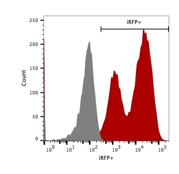
Flow Cytometry for iRFP: 4T1-Fluc-Neo/iRFP-Puro (red) or isotype control (4T1-Fluc-Neo; grey) cells were fixed with paraformaldehyde and analyzed by flow cytometry (20,000 events).

Bioluminescent images of a subcutaneous 4T1-Fluc-Neo tumor in an athymic mouse.
107 4T1-Fluc-Neo cells (Imanis Life catalog #CL020) were injected subcutaneously into the right flanks of female NCR athymic mice. Mice were imaged at day 0 on the day of cell implantation and at day 16 using a Perkin Elmer IVIS® Spectrum system, at 10-15 minutes post intraperitoneal injection of D-luciferin at 150 mg/kg. Tumor size was measured using calipers. Data from a representative mouse (ID#3876) is shown.
*Note: This is not an image from this cell line. 4T1-Fluc-Neo is a cell line generated from the same parental line using the same transduction protocol.