Anti-human NIS antibody clone VJ2
- 0.25 ml / Standard (REA003-STAN) $ 350
Description: Monoclonal antibody to human NIS clone VJ2
Antigen: Human sodium iodide symporter.
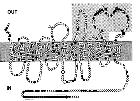
Recognizes the extracellular domains of hNIS (ref. 2). Epitope recognition by the anti-hNIS antibody clone is located between amino acid 272 and 515 located within the last three extracellular loops (shaded area).

Species: Mouse
Isotype: IgG1
Species reactivity: Human
Cross reactivity: Sheep (ref. 1). This antibody does not cross react with rat or mouse NIS
Tested Applications:
- Flow cytometry. Use at 1:50 dilution
- Immunofluorescense. Use at 1:10 to 1:50 dilution.
- This antibody VJ2 detects the native form of NIS and does not recognize the denatured form
Selected Product References:
1. Hombach-Klonisch S. Mol Cell Endocrinol. 2013; 10;367(1-2):98-108. PMID: 23291342
2. Pohlenz J. et al. J Clin Endocrinol Metab. 2000; 85 (7):2366-9. PMID: 10902780
3. Marsee DK. et al. Cancer Gene Therapy. 2004; 11, 121–127. PMID: 14730332
4. Hou P. et. al. PLoS One. 2009; 10;4(7):e6200. PMID: 19593429
5. Higuchi T. et. al. J Nucl Med. 2009; 50(7):1088-94. PMID: 19525455
6. Hou P. J Clin Endocrinol Metab. 2010;95(2):820-8. PMID: 20008023
Flow cytometry to detect human NIS expression on surface of lentiviral transduced human cell lines stably expressing human NIS
Tissue: Lentiviral transduced 293 cells expressing NIS. Type: Fresh cells Stain: Secondary antibody Alexa 555 Dilution: 1:50.

Immunofluorescence staining for human NIS expression:
Tissue: Lentiviral transduced Mel624-hNIS-Neo cells stably expressing NIS, 200xType: Cytospin of cells on glass slide. Stain: Secondary antibody Alexa 555 with Hoechst 33342. Dilution: 1:50
Tissue: Cryosection of tumor infected with an oncolytic virus expressing NIS, showing areas of NIS expressing cells, 200x. Type: OCT cryosection. Stain: Secondary antibody Alexa 555 with Hoechst 33342. Dilution: 1:50


