Anti-human NIS antibody SJ1
- 0.25 ml / Standard (REA004-STAN) $ 450
Description: Rabbit polyclonal antibody to human NIS (hNIS)
Antigen: MBP/C-terminal portion of the human NIS (amino acid 468-643) fusion protein (MBP-hNIS).
Reference: Jhiang SM. Endocrinology. 1998; 139(10):4416-9. PMID:9751526
Species: Rabbit
Reactivity: Human, Rhesus macaque
Isotype: Not applicable
Tested Applications:
- Western blot (1:3000 to 1:5000)
- Immunofluorescence (1:500 to 1:3000)
- Immunohistochemistry (1:2000 to 1:5000)
- Flow cytometry analysis (1:2000 to 1:3000)
 Lane 1: Total membrane protein from Mel624-hNIS-Neo cells (20 ug). Lane 2: Total membrane protein from control murine CT26.WT-mNIS cells (20 ug). Anti-hNIS Ab was used at 1:2000 dilution. The expected size of hyperglycosylated hNIS is 75-90 kDa (top band) and hypoglycosylated hNIS is 60-65 kDa (bottom band).

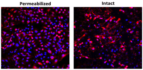
Intact or permeabilized Mel624-hNIS-Neo cells were stained with anti hNIS SJ1 antibody (1:500 dilution), followed by incubation of Alexa 594 conjugated anti-rabbit antibody.
Immunohistochemical staining:

NIS expression in (A) human thyroid or (B) VSV-mIFNβ-NIS virus treated mouse tumor xenograft. Antigen retrieval was performed on paraffin embedded sections prior to incubation with anti hNIS SJ1 antibody (1:2000). Counterstain is hematoxylin.
Flow cytometry:

Human thyroid cell, SW579 were fixed and stained with anti hNIS SJ1 (1:2000), 1h on ice. Cells were then stained with Alexa 594 conjugated anti-rabbit antibody for 30 min on ice.
 Lane 1: Total membrane protein from Mel624-hNIS-Neo cells (20 ug). Lane 2: Total membrane protein from control murine CT26.WT-mNIS cells (20 ug). Anti-hNIS Ab was used at 1:2000 dilution. The expected size of hyperglycosylated hNIS is 75-90 kDa (top band) and hypoglycosylated hNIS is 60-65 kDa (bottom band).
