Anti-human (KELE) NIS antibody (affinity purified)
- 100 ul / Standard (REA010-STAN) $ 495
Description: Affinity purified rabbit polyclonal antibody to human (KELE) NIS
Antigen: Synthetic peptide KELEGAGSWTPCVGHD corresponding to residues 618-633 of the human sodium iodide symporter, hNIS (ref.1).
Detects both the native and denatured forms of human NIS. This antibody recognizes an intracellular C-terminal epitope.
This antibody does not detect mouse, or rat NIS.
Host Species: Rabbit
Isotype: Not applicable. Polyclonal affinity purified antibody.
Concentration: 0.4 mg/mL
Species reactivity: Human
Tested Applications (see references below):
- Immunofluorescence (1:500 dilution) Ref: 2
- Immunohistochemistry (1:5,000 dilution) Refs: 3, 4
- Western Blotting (1:1,000 to 1:5,000 dilution) Ref: 2
Selected Product References:
1. Carrasco et al. Methods Enzymol. 1986. 125: 453-467.
2. Dohan et al. Mol Endocrinol 2006. 20: 1121-1137.
3. Dohan et al. J Clin Endocrinol. Metab. 2001. 86: 2697-2700.
4. Tazebay et al. Nat. Med. 2000. 6(8): 871-878.
Immunohistochemistry staining for human NIS expression (performed by Dr. Nancy Carrasco's laboratory in Dohan et al. 2001):
Thyroid tissue sections were deparaffinized. Slides were subjected to antigen retrieval using 10% citrate buffer. Slides were incubated with 1 ug/ml anti-human NIS antibody for 1h. All slides were counterstained with hematoxylin. A. normal thyroid. C. Graves' tissue. F. papillary carcinoma. K. follicular carcinoma.

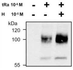
Stimulation of NIS protein expression and plasma membrane targeting by all-trans-retinoic acid (tRa) and hydrocortisone was determined by Dohan et al. 2006.
Immunoblotting for human NIS protein:
MCF-7 human breast cancer cells were treated for 48h with trans-retinoic acid (tRa) or hydrocortisone (H). Cells were lyzed and subjected to immunoblot analysis with 4nM anti-hNIS antibody. A 100 kDa band of mature NIS and 60 kDa band of partially glycoslyated polypeptide were detected.

Immunofluorescence:
NIS targeting to plasma membrane was assessed by immunoblotting with anti-hNIS antibody. Cells were permeabilized with 0.2% BSA, 0.1% triton X-100 in PBS for 10min and then quenched with 50 mM NH4Cl in PBS for 10 min. Cells were incubated with 4nM anti-hNIS antibody for 1h at room temperature. Cells were washed and incubated with anti-rabbit FITC conjugated secondary antibody. Nontreated MCF-7 (G); intracellular and faint plasma membrane-localized NIS expression in tRa-treated MCF-7 (H); clear plasma membrane localization of NIS in tRaH-treated MCF-7 (I).


